


What is a mole check and what does It Involve?
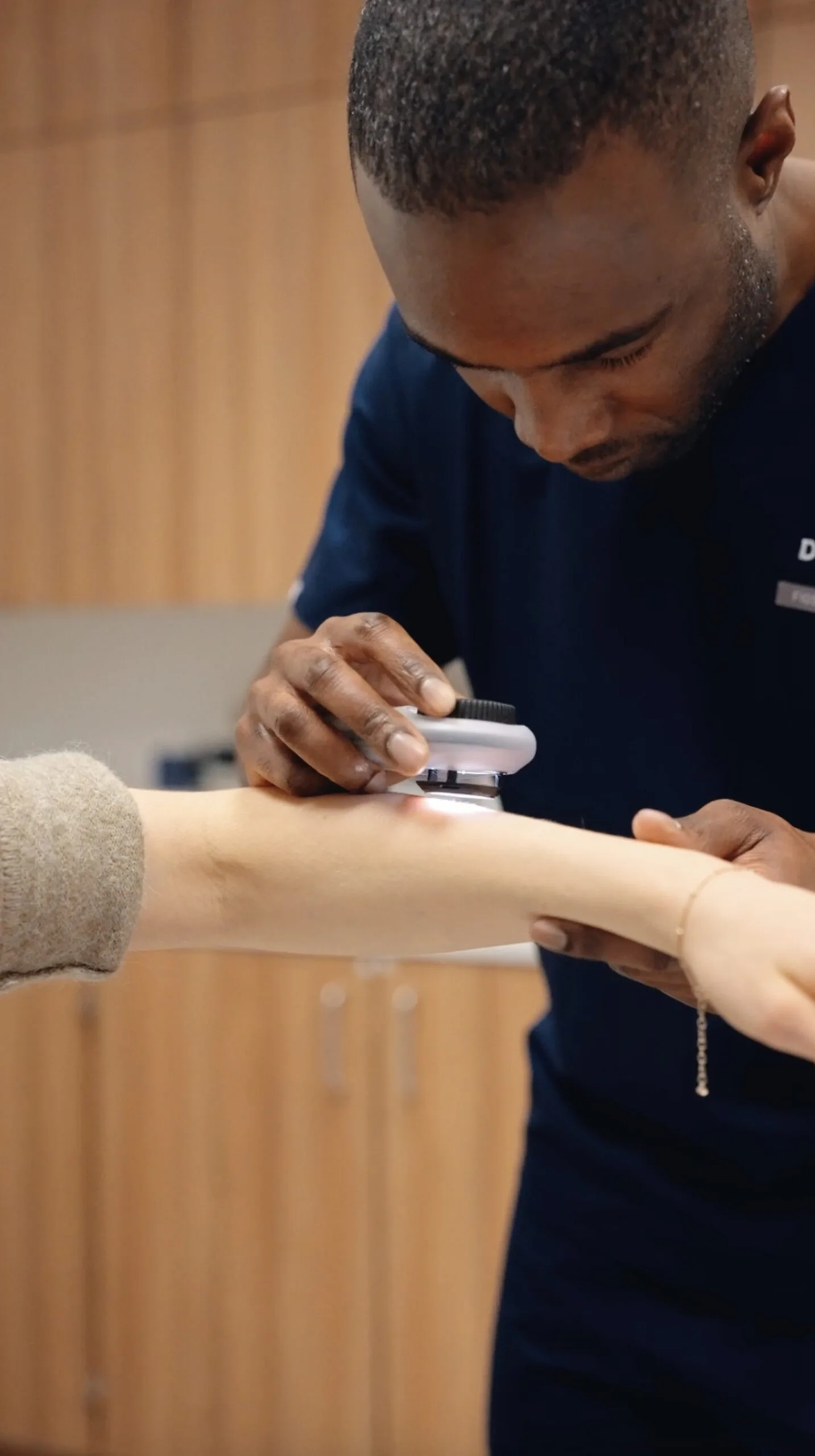
A mole check is a thorough examination of the skin by a Consultant Dermatologist to assess moles and other skin lesions for features that may require further investigation. Dermoscopy forms an important part of the assessment, allowing structures beneath the skin surface to be examined in greater detail than with the naked eye alone and improving diagnostic accuracy.
Dr Phillips examines the skin systematically and assesses any concerning or unusual lesions using established clinical and dermoscopic criteria. If a lesion requires further assessment, he will explain the findings and discuss the most appropriate next step. This may include reassurance, digital monitoring, biopsy or surgical removal.
What Is mole mapping?
For patients with multiple moles, atypical moles, or an increased risk of melanoma, VECTRA WB360 mole mapping provides a structured way to monitor the skin over time. High-resolution total-body photography creates a detailed baseline record of the skin surface, while integrated software can help identify new or changing lesions when images are compared at future visits.
The main value of mole mapping is longitudinal surveillance. Subtle changes in a mole may be easier to recognise when current images are compared directly with previous photographs rather than assessed at a single point in time.
Mole mapping is used alongside, rather than instead of, a Consultant Dermatologist’s clinical and dermoscopic assessment. It can be particularly useful for patients with large numbers of moles, atypical moles, or a personal or family history of melanoma.

Who should have a skin check?
Anyone with a new, changing or unusual mole or skin lesion
Patients with numerous moles or atypical (dysplastic) moles
Anyone with a personal or family history of melanoma or other skin cancers
Patients with significant cumulative sun exposure or a history of blistering sunburn
Organ transplant recipients or others with reduced immune function
Anyone who would like reassurance or a baseline assessment of their skin
The ABCDE warning signs
A
Asymmetry
One half differs from the other
B
Border
Irregular, blurred or poorly defined edges
C
Colour
Variation in colour within the same lesion
D
Diameter
Particularly if larger than 6 mm, although melanomas may be smaller
E
Evolution
Any change in size, shape, colour or symptoms
Types of skin cancer Dr Phillips screens for
Does skin cancer affect darker skin tones?
Skin cancer is less common in darker skin tones, particularly in black and brown skin, but it can still occur. When it does, it may affect less typical sites that are not usually associated with sun exposure.
In darker skin, melanoma can occur on areas such as the palms, soles, beneath the nails (subungual melanoma) and, more rarely, on mucosal surfaces. These presentations can be easier to overlook because they differ from the patterns more commonly seen in fair skin.
Dr Phillips has extensive experience assessing skin lesions across a wide range of skin tones and is familiar with the different ways in which skin cancer can present in black and brown skin.
What to expect at your appointment
Your consultation begins with a review of your medical history, skin cancer risk factors and any moles or skin lesions that concern you. Dr Phillips then performs a systematic examination of the skin, using dermoscopy to assess any lesions that require closer inspection.
Where appropriate, total-body mole mapping with the VECTRA WB360 system may be recommended to create a photographic baseline for future comparison. Dr Phillips will explain the findings and discuss whether any lesions require reassurance, monitoring, biopsy or removal.
Review and risk assessment
Discuss your medical history, skin cancer risk factors and any moles or lesions that concern you.
Skin and mole examination
Dr Phillips systematically examines your skin, using dermoscopy to assess any lesions requiring closer inspection.
Mole mapping where appropriate
VECTRA WB360 total-body mole mapping may be recommended to create a photographic baseline for future comparison.
Findings and next steps
Review the findings and discuss whether any lesions require reassurance, monitoring, biopsy or removal.
Mole removal in London
If a mole or skin lesion is suspicious or requires confirmation of the diagnosis, Dr Phillips may recommend a biopsy or surgical removal. The tissue is then sent for histological analysis, where it is examined under a microscope to establish a definitive diagnosis.
Benign moles can also be removed where they are symptomatic, repeatedly irritated or causing cosmetic concern. The most appropriate removal technique depends on the type, size and location of the lesion and will be discussed at consultation.
Book a consultation with Dr Derrick Phillips to have a mole assessed, mapped or removed.
Why choose Dr Derrick Phillips for skin cancer screening in London
Effective skin cancer screening relies on clinical experience and dermoscopic assessment as well as technology. As a Consultant Dermatologist with 12 years of NHS clinical experience, Dr Phillips combines systematic skin examination with dermoscopy and, where appropriate, total-body mole mapping using the VECTRA WB360 system.
Each assessment is tailored to your individual risk factors, with clear advice on whether a lesion can be reassured, should be monitored or requires further investigation or treatment. Dr Phillips also has extensive experience assessing skin lesions across a wide range of skin tones, including presentations that may look different from those typically shown in standard textbook images.

What to know about mole mapping and skin cancer screening
Early detection is an important part of skin cancer management, and a professional skin examination can identify concerning features that may not be apparent during self-examination.
Dermoscopy allows structures beneath the skin surface to be examined in greater detail than with the naked eye alone and improves diagnostic accuracy.
Mole mapping with the VECTRA WB360 system allows new and changing lesions to be identified over time and can be particularly useful for patients at increased risk of melanoma.
Skin cancer can affect all skin tones. Although it is less common in darker skin, it may occur in less typical locations and can be diagnosed at a later stage.
FAQs
Explore knowledge base
Explore expert guidance on mole checks, skin cancer awareness and looking after your skin.